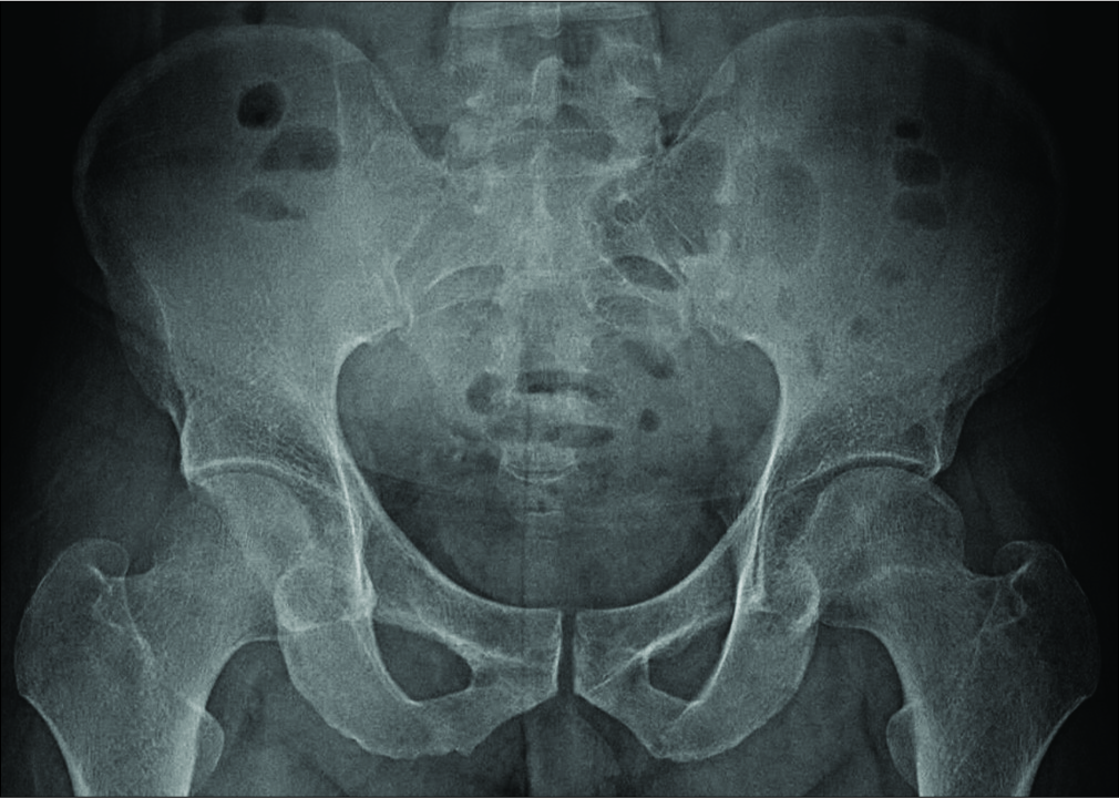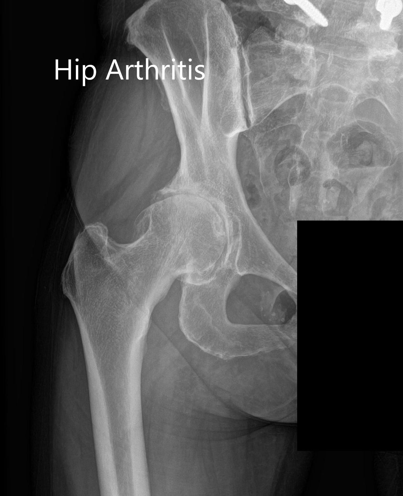

It should be borne in mind that the x-ray of the pelvic and hip joints in young children does not show the exact shape of the joint structures, since their main tissue is cartilage, which the x-rays do not display. If an X-ray of the hip joints is performed according to Launstein (Lauenstein), then the patient’s position looks like this: lying on his back, one leg in the knee bends (at an angle of 30, 45 or 90 °), while its foot rests on the shin of a straightened leg the hip of the bent limb is pulled aside as far as possible so that the hip joint takes the position of external rotation (that is, the head of the femur rotates in the acetabulum). Thus, either both kneecaps should be directed forward, or the lower limbs should be turned inward by 15-20 ° in order to adapt the femoral anti -version on radiographs of the anteroposterior femur. If radiographs of the anteroposterior thigh are made while lying on your back, one of the most common errors is image distortion when you turn the hips outside. The anteroposterior x-ray of the thigh includes images of both sides of the thigh on the same film and protrudes in the direction of the middle of the line connecting the upper part of the symphysis pubis and the anteroposterior iliac spine the distance between the x-ray tube and the film should be 1.2 meters. With conventional radiography, an anteroposterior and lateral radiography of the thigh is usually done. Also, a picture can be taken with a lateral projection, that is, the patient should lie on his side, bending his leg in the knee and hip joints. The maximum visual information is given by the x-ray of the hip joint in two projections: in the direct projection (or frontal) obtained by focusing the x-ray tube perpendicular to the body plane - front or rear, and axial (transverse or horizontal plane), fixing the elements of the joint from top to bottom - along the femur. If in the first case the procedure lasts about 10 minutes, and the picture is taken on film, then in the second method the time is halved, and the image can be in two formats, including digital. The standardized radiography technique is little dependent on the method used - analog or digital. In addition, x-rays can be prescribed for pain in the hip joint in children of different ages. The main pathology is congenital dislocation of the hip. This means that the images obtained of all articular elements have no anatomical abnormalities, for more details see - Hip JointĪn X-ray of the hip joints in children is carried out according to strict indications - only after the child reaches nine months. If the above diseases and conditions are absent, the protocol (description) of the x-ray image will indicate that the x-ray is normal. In principle, the patient's complaints about the felt pain in the hip joint are considered sufficient reason for the appointment of radiography - to establish their exact cause.

osteoarthritis, osteomyelitis and osteochondromatosis.coxitis (inflammation of the hip joint).arthritis, arthrosis of the hip joint, deforming arthrosis or coxarthrosis.juvenile epiphysiolitis of the femoral head.

congenital dislocation or dysplasia of the hip joints.traumatic damage to the hip area, in particular, femoral neck fractures.The most common indications for X-ray diagnostics of the hip joints relate to: Directing the patient to radiography, a traumatologist, orthopedic surgeon, surgeon or rheumatologist are able to assess the state of structures of this bone joint.


 0 kommentar(er)
