
The vestibulo-ocular reflex initiates when the head turns and activates the semicircular canals. Via the vestibulo-ocular reflex, the eyes move in the opposite direction of the head motion and stabilize the image on the retina. The connection between the vestibular system and extraocular muscles allows the eyes to maintain stability by fixating on a target point during head movements. The sternocleidomastoid muscle (SCM) is one of the main muscles innervated by the medial vestibulospinal tract, and a patient with the lesion tract cannot turn his or her head to the contralateral side. These neurons are responsible for the contraction of neck muscles and stabilization of head position when they receive information from the semicircular canals. They project bilaterally through the medial longitudinal fasciculus (MLF) and synapse with the medial ventral horn of the cervical cord. The neurons that travel down the medial vestibulospinal tract originate in the medial vestibular nucleus. This lateral pathway facilitates the contraction of the forelimb and hindlimb muscles to maintain balance and upright posture. Collateral nerves arise from the lateral vestibulospinal tract and synapse with the medial motoneuron, which innervates proximal muscles of the limb, in the ventral gray horn. The lateral vestibulospinal tract travels along the ventral regions of the white matter of the entire spinal cord. The lateral and inferior vestibular nuclei project motor neuron fibers that descend ipsilaterally through the lateral vestibulospinal tract. Because the spinal tract and nucleus of CN V are located close to the vestibular nuclei at the rostral medulla, a lesion of the vestibular nuclei at the level of the rostral medulla may also involve these structures leading to facial numbness and tingling.

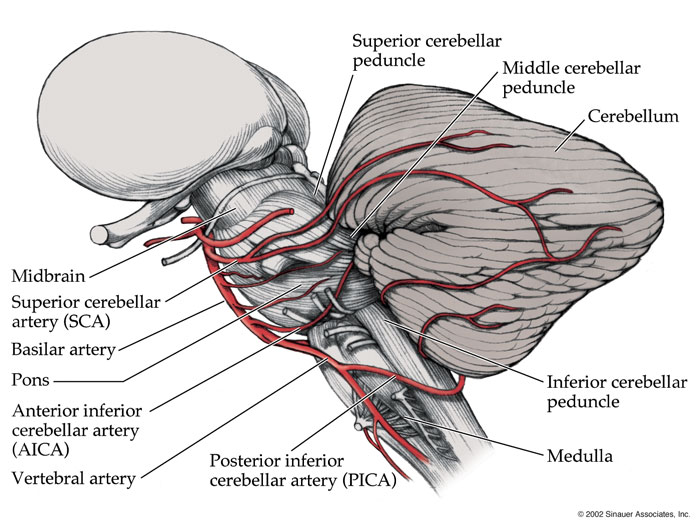
Lesions to these nuclei may lead to nystagmus, vertigo, and unsteadiness. The vestibular nuclei receive vestibular information from the CN VIII and send projections to the spinal cord, extraocular motor nuclei, thalamus, or cerebellum. They are observable near the level of the fourth ventricle in the medulla and pons. įour vestibular nuclei are lateral to the sulcus limitans and medial to the inferior cerebellar peduncle. As the vestibular portion of the cranial nerve VIII (CN VIII), the central processes of these bipolar neurons enter the brainstem at the pontomedullary junction and project to the vestibular nuclei or the flocculonodular lobes of the cerebellum via the inferior cerebellar peduncle. Hair cells of the peripheral vestibular structures receive their innervation from peripheral processes of bipolar neurons of the vestibular ganglion (also known as Scarpa’s ganglion), which is located inside the internal auditory meatus of the petrous temporal bone. It initiates a relay of sensory information through the vestibular pathways.

Upon either linear or rotational acceleration, a corresponding deflection of the hair cell bundle leads to a depolarization of the hair cell membrane potential. Their hair cells are located in the crista ampullaris, a cone-shaped neuroepithelial structure in the semicircular ducts. The semicircular ducts contain sensory receptors for dynamic equilibrium, which maintains the head position in response to the body's rotational acceleration (i.e., turning). The semicircular ducts are positioned on three perpendicular planes and correspond to three directions of the head movement. The hair cells of the utricle and saccule have stereocilia that are embedded in the otolithic membrane. The utricle and saccule contain sensory receptors for static equilibrium, which maintains the head position in response to linear acceleration of the body (i.e., starting to walk or stopping). Many afferent nerve signals originate in these peripheral vestibular organs and travel to the vestibular centers located in the brain. The utricle and saccule are also known as the otolith organs.

The membranous labyrinth comprises the utricle and saccule, three semicircular ducts, and the cochlear duct. Within the bony labyrinth, the membranous labyrinth filled with endolymph is observable. The three canals are superior, lateral, and posterior semicircular canals located posterior and superior to the vestibule. The bony labyrinth in each ear is composed of the vestibule, three semicircular canals, and the cochlea filled with perilymph. The inner ear located within the petrous temporal bone comprises the bony and membranous labyrinths.


 0 kommentar(er)
